|
||
|
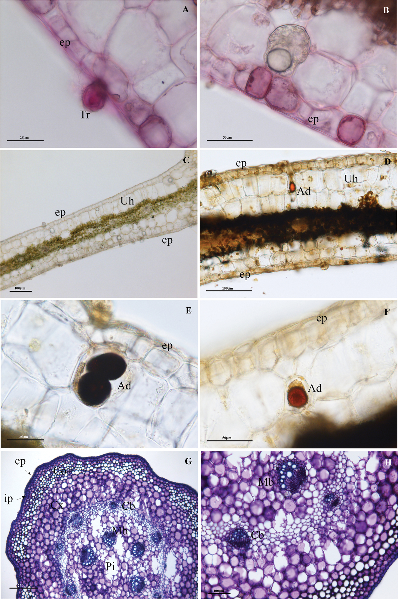
LM images of the cross-sections of P. malgassicum leaf (A–F) and stem (G, H) A PAS positivity of trichomes and epidermis cells B PAS positivity of the epidermal cells and terpenic droplet below the first layer of cells C staining with FeCl3 D leaf lamina positive to Wagner staining E detail of D showing alkaloid droplets in the hypodermis cell F detail of D showing alkaloid droplets in hypodermis idioblasts G stem stained with Toluidine blue showing the thick layer of collenchyma beneath the epidermis and the two concentric circles of vascular bundles H detail of G showing the wall thickenings of the angular collenchyma. Ad: Alkaloid droplet; Cb: cortical circle of vascular bundles; Co: angular collenchyma; cx: cortex; ep: epidermis; hp: hypodermis; Mb: medullary circle of vascular bundles; Pi: pith; Td: terpenic droplet; Tr: trichome; Uh: upper hypodermis. |