|
||
|
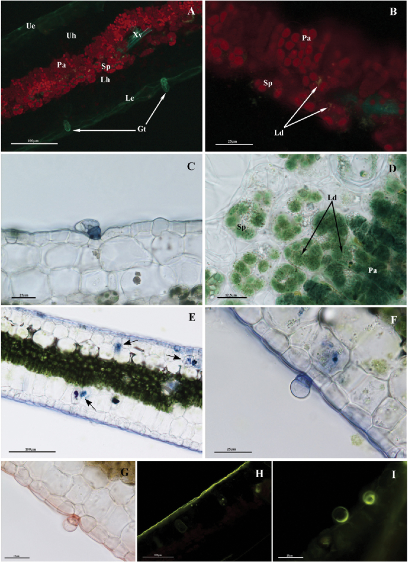
A portion of the leaf lamina in autofluorescence under blue-violet light showing the red fluorescence of chlorophyll and the red fluorescence of the trichome contents B detail of image A. Lipid droplets show green fluorescence C trichomes and cuticles appearing Sudan Black positive D sudan Black stained lipid droplets positively in the spaces between chlorenchyma cells, both in the spongy and the palisade parenchyma (black arrows) E leaf stained with NADI reaction. Positive cells can be observed in the mesophyll (arrows) F lower epidermis and trichome stained with NADI G LM images showing the trichome and cuticle slightly reddish while cutin and suberin resulted stainedbrownish with Sudan III–IV H Fluorescence images with Fluorol Yellow staining revealing the lipids in the cuticleand aggregates in the epidermis and mesophyll I detail of H): trichome stained with FY088. Ld: lipid droplet; Ue: upper/adaxial epidermis, Uh: upper hypodermis, Pa: palisade tissue, Sp: spongy tissue, Lh: lower hypodermis, Le: lower/abaxial epidermis, Gt: glandular trichomes. |