|
||
|
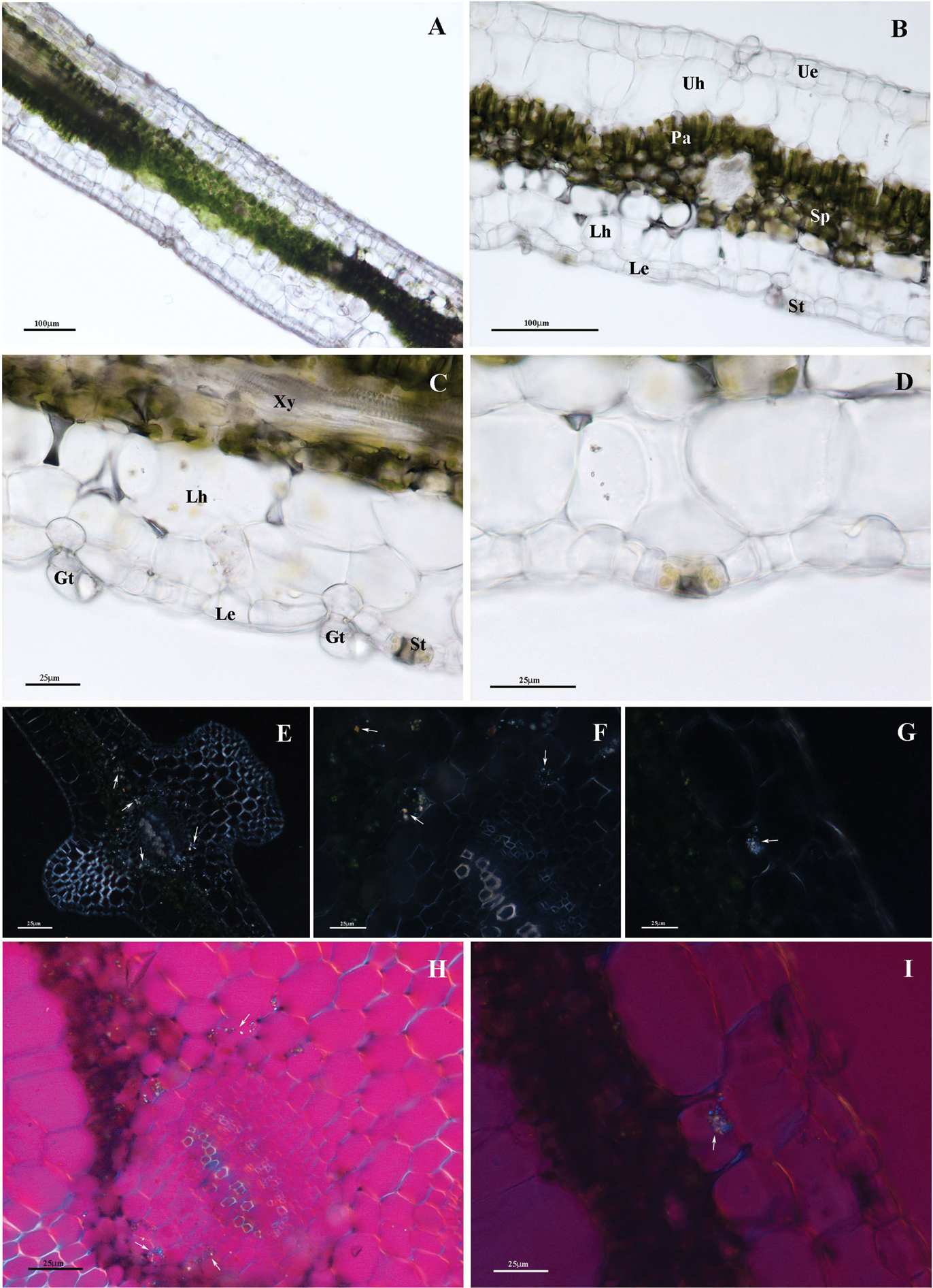
Cross-sections of P. malgassicum leaf, LM images A–D are cross-section of P. malgassicum leaf F, G LM observations with polarized light H, I LM observations using birefringence filter A portion of the leaf lamina B detail of image A C detail of image B D detail of C with stomata E portion of the leaf lamina. Presence of CaOx crystals (white arrows) within cells as well as embedded in vascular bundlecell walls F cross-section of the leaf through midrib. Abundance of CaOx crystals (white arrows) close to the bundles and embedded in the cell walls of xylemelements G presence of CaOx crystals (white arrows) in the lower hypodermis H portion of the lamina. CaOx crystals (white arrows) in the lower hypodermis I cross-section of the leaf through midrib showing abundant CaOx crystals (white arrow) close to the bundles and embedded in the cell walls of xylemelements. Xy: xylem, Gt: glandular trichomes; Ue: upper/adaxial epidermis, Uh: upper hypodermis, Pa: palisade tissue, Sp: spongy tissue, Lh: lower hypodermis, Le: lower/abaxial epidermis, St: stomata. |